
Fibrous Dysplasia
A benign bone disorder in which normal bone is replaced by fibrous tissue and immature woven bone, creating a distinctive "ground glass" appearance on X-ray. Most patients have an excellent long-term outlook with observation, bisphosphonates or targeted surgery when needed.
What is fibrous dysplasia?
Fibrous dysplasia (FD) is a benign, non-inherited disorder of bone development. Normal bone and marrow are gradually replaced by fibrous tissue mixed with irregular islands of woven bone, giving the classic ground-glass appearanceon X-ray. It typically becomes apparent between 5 and 30 years of age and accounts for around 5–7% of all benign bone tumours.1,2
Who gets fibrous dysplasia and where does it occur?
Most lesions apparent by age 5; symptoms present in childhood or adolescence
Of all benign bone tumours
Followed by tibia, humerus, pelvis, ribs and craniofacial bones
- • Milder form — often silent and found by chance
- • Better prognosis; usually stabilises after skeletal maturity
- • Makes up 85–90% of all cases
- • More severe; can progress into adulthood
- • May be part of McCune-Albright syndrome (FD + café-au-lait spots + precocious puberty)
- • May be part of Mazabraud syndrome (FD + soft-tissue myxomas)
What causes fibrous dysplasia?
Fibrous dysplasia is caused by a somatic mutation in the GNAS gene on chromosome 20 — meaning it happens during early development and is not inherited and cannot be passed to children.
- • Increases cyclic AMP (cAMP) inside bone-forming cells
- • Blocks normal maturation of skeletal stem cells
- • Produces immature, disorganised woven bone in place of normal bone
What are the symptoms of fibrous dysplasia?
Polyostotic disease can additionally cause craniofacial asymmetry, hearing or vision changes and, in McCune-Albright syndrome, hormonal problems such as precocious puberty or hyperthyroidism.1,6
How is fibrous dysplasia diagnosed?
- • Plain X-ray: 'ground glass' matrix with a well-defined lesion — usually diagnostic on its own for typical monostotic disease
- • Bone scan: assesses activity and screens for other affected bones in polyostotic disease
- • CT: best detail of bone architecture, especially in the skull base and pelvis
- • MRI: reserved for atypical lesions or when a soft-tissue mass is suspected
- • Only for doubtful cases — typical X-ray appearance is usually enough
- • Not routinely required for classic monostotic lesions
- • Mandatory if malignant transformation is suspected
- • Performed by an orthopaedic oncology team as an image-guided core biopsy
Understanding benign bone tumours
A short explainer on how we approach benign bone lesions like fibrous dysplasia — diagnosis, when treatment is needed, and what recovery looks like.
Current treatment options for fibrous dysplasia
Most patients need only observation. Treatment is tailored to the location, symptoms, fracture risk, deformity and whether the disease is monostotic or polyostotic. The aim is the least invasive plan that will reliably relieve pain and protect the bone.1,6
Conservative observation
- • Best for: asymptomatic, stable, monostotic lesions
- • Plan: follow-up X-rays every 6 months for stable disease
- • No intervention needed for inactive lesions
- • Watch for new symptoms, activity change or rapid growth
Bisphosphonate therapy (symptomatic disease)
- • Reduces bone turnover markers (alkaline phosphatase)
- • Radiological strengthening of involved bone
- • Typically limited to about 2 years of therapy
Surgery (when conservative care isn't enough)
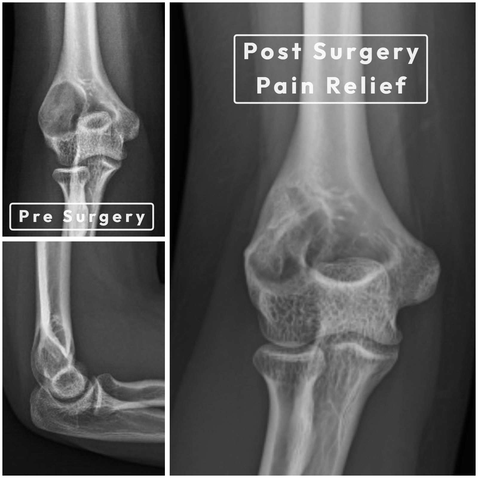
Considered for progressive pain despite medical therapy, significant functional impairment, deformity correction, impending fractures (especially around the hip) or established pathological fractures.1,7
- • Extended curettage with a high-speed burr
- • Bone cement filling — excellent functional scores (MSTS ~27/30) for contained lesions
- • Bone grafting alone has higher resorption rates and is less durable
- • Preferred for long-bone fractures and deformity correction
- • PFNA (proximal femoral nail) for proximal femur disease
- • Intramedullary nailing for diaphyseal lesions
- • ~85% revision-free survival at 2 years
What is the outlook with fibrous dysplasia?
The outlook is excellent for most patients. Monostotic disease usually stabilises after skeletal maturity, symptomatic patients respond well to bisphosphonates, and targeted surgery preserves function even in severe disease. A small subset with polyostotic disease need lifelong multidisciplinary care involving orthopaedics, endocrinology and, sometimes, genetics.1,4,6
Can fibrous dysplasia become cancerous?
Malignant transformation is rare (0.4–4%) and usually occurs after the fourth decade of life. It does not require prior radiotherapy to happen. The commonest cancers arising from long-standing FD are osteosarcoma (~70%), fibrosarcoma (~20%) and chondrosarcoma (~10%).9
- • Rapidly increasing pain, particularly at rest or at night, without any injury
- • New destructive changes on imaging, cortical breakthrough or a soft-tissue mass
- • A new osteolytic lesion within previously stable FD
- • Any of the above appearing after age 40
Which approach is best for me?
- • The lesion is asymptomatic and found by chance
- • It is monostotic and away from a weight-bearing hot spot
- • There is no deformity or fracture risk
- • Persistent bone pain despite simple analgesia
- • Polyostotic disease with multiple painful lesions
- • As part of a wider plan alongside monitoring
- • Impending or established pathological fracture
- • Progressive deformity such as shepherd's crook of the femur
- • Pain and functional impairment not controlled by medicines
Long-term monitoring plan
- • 6-monthly clinical and X-ray review for stable lesions
- • Endocrine evaluation for all patients with polyostotic disease
- • Multidisciplinary follow-up for McCune-Albright syndrome
- • Life-long surveillance for polyostotic and syndromic disease
- • Sudden severe pain or a suspected fracture
- • Rapid worsening of bone pain without any injury
- • New neurological symptoms such as vision or hearing changes
- • Any change in function that affects daily activities
Next Steps — Talk to a Specialist
References
- 1NCBI PMC — Fibrous dysplasia of bone: a review of current management. Journal of Clinical Orthopaedics and Trauma, 2018.
- 2Academic.oup — Fibrous dysplasia / McCune-Albright syndrome: clinical and translational perspectives. Endocrine Reviews, 2020.
- 3PubMed — Bisphosphonate treatment in fibrous dysplasia: significant clinical response. Clin Orthop Relat Res, 2001.
- 4Wiley Online Library — Long-term outcome of bisphosphonate treatment in fibrous dysplasia / McCune-Albright syndrome. J Bone Miner Res, 2017.
- 5AJR — Fibrous dysplasia of bone: radiographic follow-up of 138 patients. American Journal of Roentgenology, 2020.
- 6PubMed — Best practice management guidelines for fibrous dysplasia / McCune-Albright syndrome. Orphanet Journal of Rare Diseases, 2019.
- 7PubMed — Long-term results of proximal femoral fibrous dysplasia treatment. J Bone Joint Surg Am, 1998.
- 8NCBI PMC — Craniofacial fibrous dysplasia: clinical guidelines for management. 2012.
- 9NCBI PMC — Malignant transformation in fibrous dysplasia of bone: a systematic review. 2018.
Important Notice: This information is provided for educational purposes only and is not a substitute for professional medical advice, diagnosis, or treatment. The content on this page should not be used to diagnose or treat any health condition.
Always seek the advice of a qualified orthopaedic oncologist or healthcare provider with any questions about fibrous dysplasia or any other medical condition.
Treatment decisions are individualised and depend on disease type (monostotic vs polyostotic), location, symptoms, patient age, associated conditions and response to conservative therapy. Only a qualified specialist can determine the most appropriate treatment approach for your situation.
Multidisciplinary care may be needed, particularly for polyostotic disease or McCune-Albright syndrome, involving orthopaedic oncologists, endocrinologists, geneticists and other specialists.
In case of emergency — severe pain, suspected fracture, neurological changes or vision/hearing problems — seek immediate medical attention or contact your local emergency services.
The Orthoncology Clinic is committed to providing current, evidence-based information while emphasising the importance of professional medical consultation for all health-related decisions.