Non-Ossifying Fibroma
A very common, harmless fibrous defect in a child's bone that is usually found by accident on an X-ray and, in most children, heals completely on its own without any treatment.
What is a non-ossifying fibroma?
A non-ossifying fibroma (NOF) is a benign (non-cancerous) fibrous defect that develops in the outer shell (cortex) of a growing long bone. It is one of the commonest bone lesions of childhood — present in roughly 20–30% of children aged 4–10 years. NOFs are best thought of as developmental defects of bone rather than true tumours.1,7
Who gets NOF and where does it occur?
Peak incidence; may be diagnosed up to age 20
Boys affected about twice as often as girls
Distal femur, proximal and distal tibia, fibula
What causes a non-ossifying fibroma?
The exact cause is unknown. The most widely accepted explanation is a developmental defect that appears while the bone is growing, where a small area of cortex is filled with fibrous tissue instead of normal bone.1
- • Not caused by injury, diet or activity
- • No known genetic or environmental risk factors
- • Nothing a parent did or failed to do
- • Multiple NOFs with skin angiomas suggest the very rare Jaffe-Campanacci syndrome
What symptoms should I look out for?
The majority of NOFs cause no symptoms at all and are discovered by chance on an X-ray taken for another reason.2
- • New or worsening pain at the lesion site
- • Pain that interferes with school, daily activity or sport
- • Swelling or a change in the shape of the affected bone
- • A suspected fracture — sudden severe pain after a minor injury
How is non-ossifying fibroma diagnosed?
- • A plain X-ray is sufficient for diagnosis in typical cases
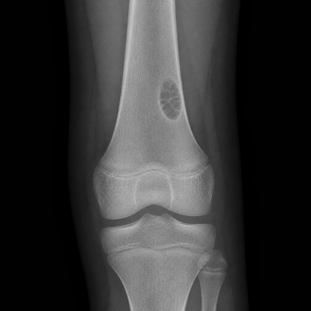
- • Well-defined, eccentric lesion centred on the cortex
- • Sclerotic margins with accentuated trabeculations
- • Homogenous matrix with a bubbly, polycyclic multiloculated pattern
- • No cortical destruction, periosteal reaction or soft-tissue mass
- • MRI is rarely required — reserved for atypical appearances or when another diagnosis is suspected
- • A confidently typical NOF is a 'leave alone' lesion and needs no further imaging
- • A biopsy is almost never necessary; when the picture is unclear, a planned biopsy resolves it safely
Ritschl staging — how an NOF evolves
Small oval lesion near the growth plate, no sclerotic border.
Larger polycyclic lesion with a thin sclerotic border — highest fracture risk.
Progressive sclerosis with ossification — the lesion is healing.
Complete healing or disappearance of the lesion.
Staging matters because it guides how closely a lesion needs to be watched. Stage B lesions progress over an average of about 21 months.2
Benign bone tumours — what patients need to know
A short explainer on how benign bone lesions such as non-ossifying fibroma are diagnosed, monitored and treated at The Orthoncology Clinic.
Watch on YouTube →How is non-ossifying fibroma treated?
For the overwhelming majority of children, the right treatment is no treatment at all — simply confirming the diagnosis and watching the lesion until it heals. In our practice, around 98% of NOFs are managed successfully by observation alone.1,2
Observation — the preferred approach
- • Most NOFs either stay stable or regress spontaneously
- • Repeat X-rays every 6 months are used only when the lesion is suspected to be enlarging, involves more than 50% of the bone diameter, or is a Stage B lesion longer than 44 mm with more than 75% bone involvement
- • Most lesions disappear by age 20–25
- • No malignant transformation has ever been reported
When surgery is genuinely needed
- • Large lesions involving >50% of bone diameter or >33 mm in length
- • Symptomatic lesions causing persistent pain
- • Pathological fracture (uncommon but well described)
- • Failure of conservative management in exceptional cases
- • Curettage with bone grafting — the traditional approach
- • Curettage with calcium sulfate pellets — a newer bone-substitute technique
- • Percutaneous cryoablation — a minimally invasive option
- • External fixation or internal fixation for large lesions at fracture risk
Pathological fractures
- • Arata criteria: lesions >50% of bone diameter or >33 mm in length carry fracture risk
- • Stage B lesions carry the highest risk
- • Tibial NOFs are more prone to fracture
- • Fractures typically occur around age 10
- • Most heal well with cast immobilisation
- • Surgery is reserved for unstable fractures or failed non-operative care
- • Healing is normal in both operative and non-operative cases
What is my child's outlook with NOF?
Which approach is right for us?
- • The X-ray appearance is typical of NOF
- • There are no symptoms or only mild activity-related ache
- • The lesion is small — under 50% of bone diameter and under 33 mm
- • The lesion is Stage B on Ritschl staging
- • It involves more than 50% of the bone diameter
- • It appears to be enlarging on serial X-rays
- • Pain persists despite conservative care
- • The lesion is large with a real fracture risk
- • A pathological fracture has occurred and the bone is unstable
When do we need follow-up?
- • Stage B lesions: review every 6–12 months until Stage C is reached
- • Large lesions (>50% bone diameter): monitor for fracture risk
- • Symptomatic children: regular review until pain-free
- • Discharge: once Stage C or D is reached with no symptoms
- • No activity restrictions for most NOFs
- • Sports participation is generally safe unless fracture risk is high
- • Pain-guided activity — a short rest if activity-related pain appears
- • Report any new symptom promptly rather than waiting for the next visit
Next Steps — Talk to a Specialist
References
- 1Imaging of Non-ossifying Fibromas: A Case Series with Natural History. NCBI PMC, 2021.
- 2Non-ossifying fibroma: natural history with emphasis on Ritschl staging and fracture risk. PubMed, 2016.
- 3Operative Treatment of a Patient with a Pathological Fracture through a Large Non-ossifying Fibroma: A Case Report. Journal of Orthopaedic Case Reports, 2024.
- 4Non-ossifying fibroma — imaging features and management. Radiopaedia, 2025.
- 5Nonossifying Fibroma — patient information. OrthoInfo (AAOS), 2022.
- 6Multiple non-ossifying fibromas: management and outcomes. ScienceDirect, 2023.
- 7Prevalence of non-ossifying fibroma in children and adolescents. NCBI PMC, 2024.
Important Notice: This information is provided for educational purposes only and is not a substitute for professional medical advice, diagnosis or treatment. It should not be used to diagnose or treat any health condition.
Always seek the advice of a qualified orthopaedic oncologist or healthcare provider with any questions about non-ossifying fibroma or any other medical condition. Never disregard professional medical advice or delay seeking it because of something you have read here.
Treatment varies with the size and location of the lesion, its Ritschl stage, the child's age, symptoms and activity level. While the great majority of NOFs are managed successfully with observation, individual cases may need a different approach.
In case of emergency — severe pain, suspected fracture or significant loss of function — seek immediate medical attention or contact your local emergency services.
The Orthoncology Clinic is committed to providing current, evidence-based information while emphasising the importance of professional medical consultation for all health-related decisions.