Osteofibrous Dysplasia & the OFD-like Adamantinoma Spectrum
A rare, benign fibro-osseous lesion of childhood that most often affects the front of the shin bone (tibia), sitting on a spectrum with OFD-like and classic adamantinoma. Careful biopsy and long-term follow-up help distinguish lesions that only need watching from those that need surgery.
What is osteofibrous dysplasia?
Osteofibrous dysplasia (OFD) is a rare, benign fibro-osseous lesion of bone. It shows woven bone trabeculae rimmed by osteoblasts within a fibrous stroma and most often affects the anterior cortex of the tibia in young children. It typically presents before age 10 and either stabilises or regresses after skeletal maturity.1,4
The OFD-like adamantinoma spectrum
Benign childhood lesion that often remodels after growth.
Intermediate lesion with focal epithelial islands on biopsy; behaves more aggressively than classic OFD.
Low-grade malignancy with prominent epithelial nests; needs wide excision and long-term surveillance.
Who gets OFD and where does it occur?
Classic OFD in early childhood; OFD-like adamantinoma peaks in adolescence
Slight male predominance
Front cortex of the shin bone; occasionally fibula
Most children present with a painless or mildly painful shin swelling. Some develop anterior bowing of the tibia leading to a leg-length difference. A pathological fracture after minor injury is uncommon but well described.1,4
What causes OFD and OFD-like adamantinoma?
The exact cause is unknown. Proposed mechanisms include a developmental error in bone formation, changes after minor trauma, or a genetic predisposition.
- • No known environmental or hereditary risk factors
- • OFD-like adamantinoma may reflect transformation of OFD over time
- • Some cases may be early adamantinomas that were undersampled at biopsy
What symptoms should raise concern?
How is OFD diagnosed and told apart from adamantinoma?
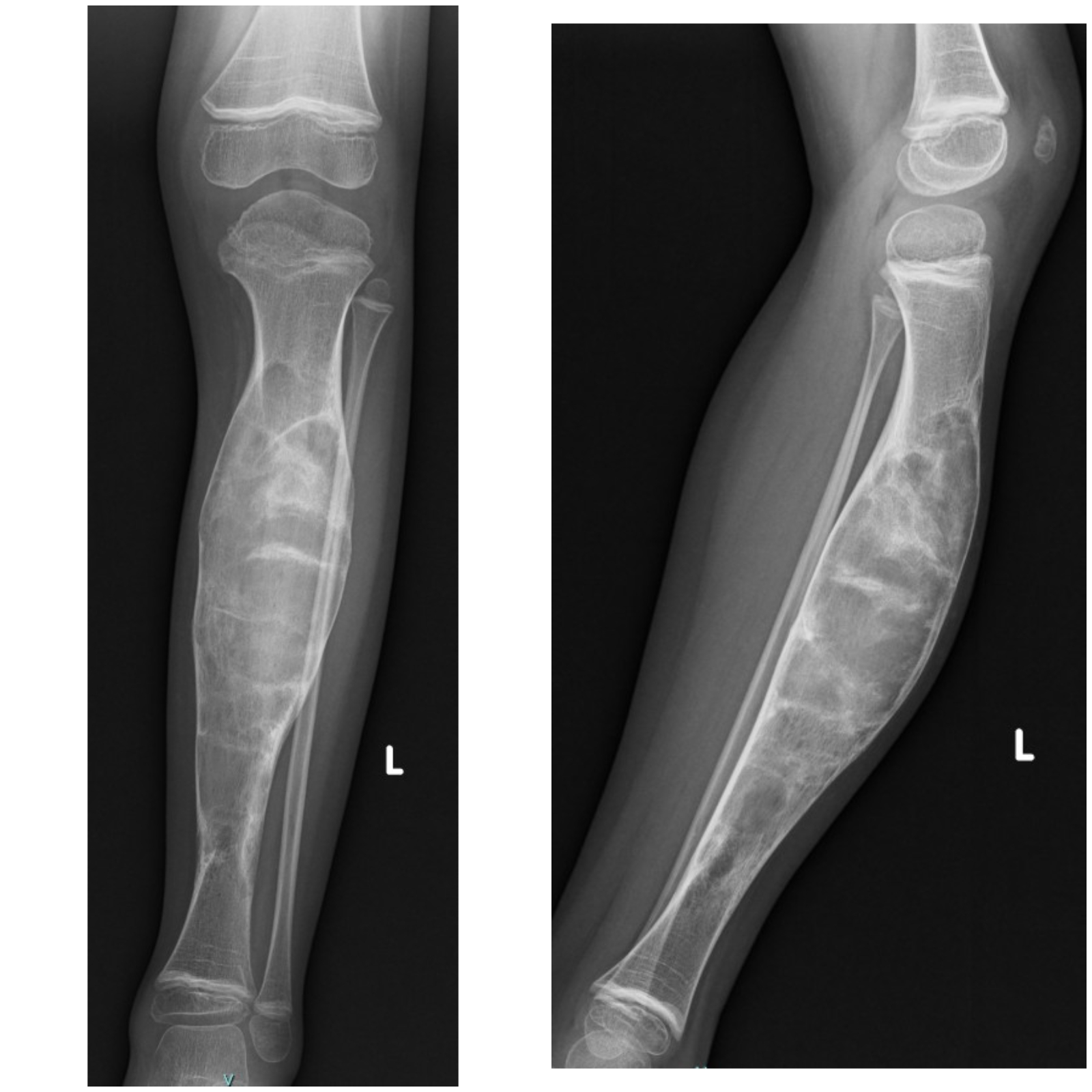
- • Plain X-ray: well-defined intracortical lytic lesion in the anterior tibia with a sclerotic rim (OFD); occasional cortical breach suggests OFD-like adamantinoma
- • MRI: assesses medullary involvement and soft-tissue extension — cortical breach is a red flag
- • CT: evaluates cortical integrity in detail
- • Whole-body imaging in selected cases to look for rare metastases
- • Classic OFD: fibrous stroma with osteoblastic rimming, no epithelial cells
- • OFD-like adamantinoma: focal epithelial cell nests staining positive for cytokeratins and CD99
- • Classic adamantinoma: prominent epithelial islands throughout the lesion
- • Sampling error is a real risk — biopsy is planned with the surgical team to preserve future limb-salvage options
Understanding benign bone tumours in children
A short explainer on how we approach benign bone lesions like OFD — the role of imaging, when a biopsy is needed and how we decide between observation and surgery.
Current treatment options along the OFD spectrum
Treatment depends on where the lesion sits on the spectrum, the child's age and skeletal maturity, symptoms and any deformity. The overarching aim is the least invasive plan that will reliably control the disease and preserve limb function.1,4
Classic OFD — observation first
- • Most OFD lesions stabilise or regress after skeletal maturity
- • Asymptomatic children need no intervention, just monitoring
- • Bracing or activity modification helps with tibial bowing
- • Surgery — usually a corrective osteotomy — is reserved for progressive deformity or functional impairment
OFD-like & classic adamantinoma — wide excision
- • Wide local excision with negative margins to prevent progression
- • Limb-salvage reconstruction planned around the child's age and remaining growth
- • Careful histological review of the whole specimen to exclude classic adamantinoma
Reconstruction after tibial resection
- • Custom tibial endoprosthesis with soft-tissue flap coverage
- • Allograft or allograft-prosthesis composite for diaphyseal defects
What is the outlook along the OFD spectrum?
Which approach is best for me?
- • Biopsy confirms classic OFD
- • The lesion is asymptomatic and stable
- • There is no significant tibial bowing or fracture risk
- • Biopsy shows OFD-like adamantinoma
- • The lesion is enlarging or painful
- • Imaging shows cortical breach or soft-tissue extension
- • Biopsy confirms classic adamantinoma
- • Reconstruction with endoprosthesis, allograft or vascularised fibula is feasible
- • Amputation reserved for tumours that cannot be safely reconstructed
Long-term monitoring plan
- • Clinical review and X-ray every 6–12 months until skeletal maturity
- • Watch for new pain, enlargement or cortical breach
- • Extended surveillance if biopsy features are borderline
Next Steps — Talk to a Specialist
References
- 1Van Rijn RR et al. — Adamantinoma: an updated review of the imaging, pathology and management. NCBI PMC, 2021.
- 2Zhang Z et al. — Epidemiological study of adamantinoma from the SEER database. NCBI PMC, 2020.
- 3Kinnunen AR et al. — Custom total tibia endoprosthesis reconstruction after resection of tibial tumours. NCBI PMC, 2018.
- 4OrthoInfo (AAOS) — Osteofibrous Dysplasia and Adamantinoma: patient information article.
- 5IJORO — Long-term outcome of tibial adamantinoma treated with wide excision and reconstruction, 2025.
- 6Journal of Orthopaedic Case Reports (JOCR) — Persistent Challenges: A Recurrent Adamantinoma Case Report, 2024.
Important Notice: This information is provided for educational purposes only and is not a substitute for professional medical advice, diagnosis, or treatment. The content on this page should not be used to diagnose or treat any health condition.
Always seek the advice of a qualified orthopaedic oncologist or healthcare provider with any questions about osteofibrous dysplasia, adamantinoma or any other medical condition.
Treatment decisions are individualised and depend on where the lesion sits on the OFD-adamantinoma spectrum, the child's age and skeletal maturity, symptoms and imaging findings. Only a qualified specialist can determine the most appropriate treatment approach for your situation.
Lifelong follow-up is essential for OFD-like and classic adamantinoma because of the potential for late recurrence and progression along the spectrum.
In case of emergency — severe pain, suspected fracture, or rapid change in a known lesion — seek immediate medical attention or contact your local emergency services.
The Orthoncology Clinic is committed to providing current, evidence-based information while emphasising the importance of professional medical consultation for all health-related decisions.