Back to Bone CancersPatient Awareness · Bone Cancers

Adamantinoma — a patient guide

Adamantinoma is a rare, slow-growing, low-grade malignant bone tumour that classically affects the tibia. Although it accounts for less than 1% of primary bone tumours, its locally aggressive behaviour and risk of very late recurrence demand expert surgery and lifelong follow-up.

>95%
5-year survival with surgery
~85%
Arise in the tibial shaft
>90%
Limb salvage achievable

What is adamantinoma?

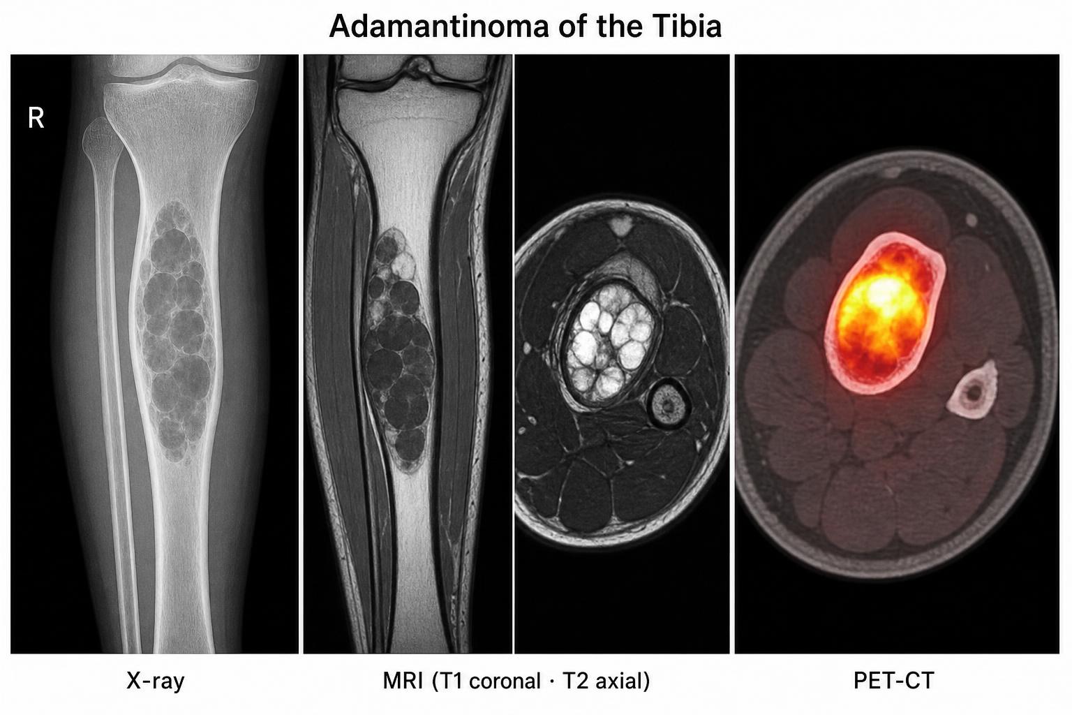
Adamantinoma is a rare bone tumour that originates from epithelial cells set within a fibrous bone background. It grows slowly but is locally aggressive and can recur many years after the original surgery. Distant spread is uncommon but possible — most often to the lungs.

Key point: A rare low-grade bone cancer where complete surgical removal is the cornerstone of cure.

Who gets adamantinoma?

  • Age: Most common in adults aged 20–50 years; rare in children and the elderly.
  • Gender: Slight male predominance.
  • Common site: Mid-shaft of the tibia in ~85% of cases; less commonly fibula, femur, ulna or humerus.
  • Origin: Believed to arise from epithelial cell remnants or from osteofibrous dysplasia.
Red flags

When to seek specialist review

Persistent shin pain

Dull ache over the front of the leg lasting weeks to months, worse at night.

Shin swelling or deformity

A slowly enlarging bony swelling or visible bowing of the tibia.

Fracture from minor trauma

A pathological fracture after a trivial fall warrants urgent imaging.

Recurrent ‘infection’

Recurrent shin symptoms mistaken for chronic osteomyelitis should be reviewed by a bone-tumour specialist.

Diagnosis

How is adamantinoma diagnosed?

A combination of imaging and an image-guided biopsy at a specialist centre confirms the diagnosis and guides treatment planning.

X-rays

Multiloculated lytic ‘soap-bubble’ lesion of the tibia with cortical thinning or disruption.

MRI

Defines tumour margins, intramedullary extent and soft-tissue involvement for surgical planning.

CT / PET-CT

CT assesses bone architecture; chest CT or PET-CT rules out the rare metastases.

Core needle biopsy

Confirms diagnosis — epithelial nests in fibrous stroma, positive for keratins and CD99.

Clinical clue: a multiloculated ‘soap-bubble’ lesion in the tibial shaft of a young adult should always raise suspicion of adamantinoma or osteofibrous dysplasia and warrants specialist review.
Adamantinoma of the tibia — imaging collage. Left: plain X-ray showing a multiloculated lytic 'soap-bubble' lesion in the tibial shaft. Middle: MRI with T1 coronal and T2 axial images showing an intramedullary lesion with cortical involvement. Right: PET-CT axial slice demonstrating intense FDG uptake in the tibial diaphysis.
Imaging of an adamantinoma of the tibia — plain radiograph showing a multiloculated lytic ‘soap-bubble’ lesion, MRI (T1 coronal and T2 axial) defining tumour extent, and PET-CT demonstrating intense metabolic uptake in the tibial shaft.
Treatment

Surgery is the primary treatment

Wide en-bloc resection with limb-preserving reconstruction is successful in the vast majority of patients with adamantinoma.

Primary treatment

Wide en-bloc resection

Surgery is the gold standard, with the aim of clear negative margins.

  • Limb salvage is achieved in over 90% of patients
  • Amputation reserved for very large or multiply recurrent tumours
  • Surgery planned at a specialist bone-tumour centre
Reconstruction

Restoring the tibia

The defect after resection is rebuilt using a combination of biological and prosthetic options.

  • Allograft or autograft bone (segmental or osteoarticular)
  • Vascularised fibula graft — especially in young patients
  • Custom megaprosthesis or endoprosthesis
  • Graft–prosthetic composites for complex defects
Adjuvant therapy

Chemotherapy & radiotherapy

Both have very limited roles in adamantinoma.

  • Chemotherapy: not routinely used
  • Radiotherapy: occasionally for unresectable or recurrent disease
  • Multidisciplinary team decision in selected cases

Rehabilitation

  • Biological grafts: 6–9 months of protected weight-bearing
  • Prosthetic reconstructions: earlier mobilisation
  • Physical therapy for strength, range of motion and function
  • Psychological support for adjustment and quality of life

Follow-up schedule

  • Every 3–4 months for the first 2 years
  • Every 6 months from year 3 to year 5
  • Annual review thereafter — for life
  • Long-term — late recurrence can occur after 8 years or more

Real-life patient experience

Case · Mid-tibial adamantinoma

Vascularised fibula reconstruction in a young man

A young man underwent extensive mid-tibial resection of an adamantinoma with reconstruction using a vascularised fibula graft. Despite the complexity of the surgery, at 5-year follow-up he demonstrated full functional recovery with no signs of disease recurrence — highlighting the importance of individualised treatment and multidisciplinary care.

Take-home summary

  • Adamantinoma is a rare, low-grade malignant bone tumour, classically of the tibia.
  • Wide surgical resection with negative margins is the gold standard treatment.
  • Limb salvage with reconstruction is possible in over 90% of patients.
  • Five-year survival exceeds 95% with complete surgical excision.
  • Local recurrence (15–30%) can be late — often beyond 8 years from surgery.
  • Lifelong, multidisciplinary follow-up is essential for best outcomes.

References

  1. Shimizu J et al. Clinical outcomes in patients with adamantinoma. J Surg Oncol, 2024.
  2. Houdek MT et al. Long-term outcomes of adamantinoma: a case series. Bone Joint J, 2021.
  3. Farooque K et al. Recurrent adamantinoma — case report. J Orthop Case Rep, 2024.
  4. OrthoInfo (AAOS). Osteofibrous dysplasia and adamantinoma, 2012.
  5. National Cancer Institute. Adamantinoma information, 2025.