
Giant cell tumour is called benign, but it quietly eats the bone that holds a joint together. In an 18-year-old cricketer, the goal was never just to remove a tumour — it was to give him back a knee he could run on.
Welcome back to Sutures & Stories.
A tumour that doesn’t discriminate
Giant cell tumour of bone is uncommon in young people, and it makes no distinction between the studious and the sporty. Bone tumours as a group are rare, and treating them well is genuinely a team effort. These lesions can turn up in a six-year-old or in someone of eighty-five. GCT is technically classified as benign — a label that undersells it. It grows, and it eats away the bone it occupies, almost always right beside a joint, weakening the very pillars that hold that joint together. Only rarely does it travel to other organs. In young patients, whose growth plates are still open and active, that behaviour is especially difficult to manage.
An 18-year-old who wanted the IPL
One such patient was an 18-year-old boy from a tier-2 city in Karnataka, who began hobbling and limping while playing cricket. He had been dreaming of training for the big leagues — the IPL among them — and this nagging problem stopped him in his tracks.
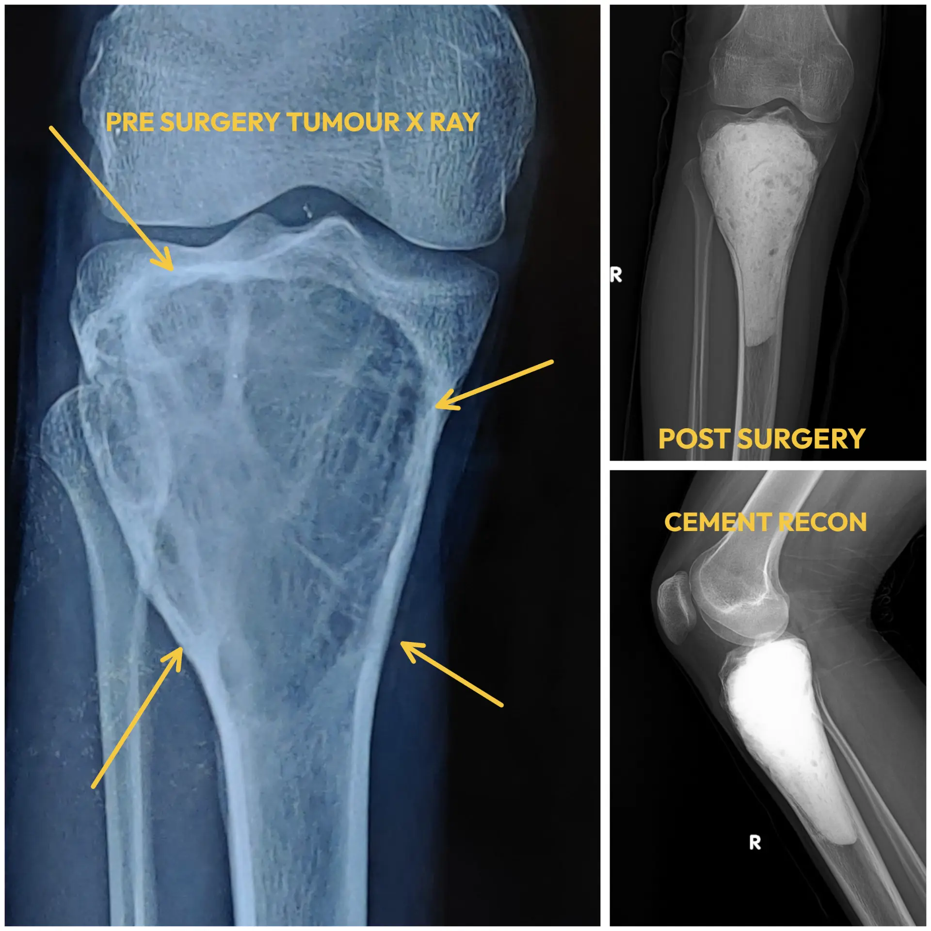
As with every suspected bone tumour, we ran the standard diagnostic work-up, which revealed an aggressive form of giant cell tumour with a marginally elevated Ki-67 index. That finding alone would not have changed the plan. But the sheer size of the lesion — and his anxiety about getting back to college, let alone back to sport — told us we needed to be aggressive in clearing the disease. That was done through extended curettage.
Grafts, cement and a COVID-era compromise
Next came the decision on reconstruction. In young patients I generally prefer bone grafts — specifically bone bank allografts. But this was during the COVID years, when bone donations had all but dried up, so I used bone cement instead. Cement carries an added advantage: the heat it releases as it sets helps neutralise any residual tumour cells. Given how much bone had been lost, I was apprehensive about his recovery — yet, surprisingly, the outer shell of bone held strong, even though the tumour had hollowed out nearly 90% of his upper tibia, right down to the diaphysis.
The follow-up that became a video
Time passed, and every follow-up visit was about two things at once: watching for any sign of relapse, and gently nudging him back towards independence. The real surprise came a year after surgery, when he sent me a video of himself playing cricket with friends. Part of me wanted to call him and his family and gently scold them for the risk — and then I reminded myself: I’m only a pair of hands, doing my best for whatever comes my way. And what greater reward is there than watching a young man who once worried about simply walking again, return to the sport he loves?
Watch the full case story of this young cricketer’s giant cell tumour of the knee — from diagnosis to his return to the game.
Watch on YouTube →This is an educational video, shared after due informed consent and with individual privacy protected.
Time and again, cases like this remind me that biology is king — and that sometimes all we need to do is push our young patients forward and encourage them to trust their bodies.
A full diagnostic protocol, including biopsy, revealed an aggressive GCT with a mildly raised Ki-67 index.
Extended curettage with cement reconstruction — the outer shell of bone held, despite ~90% internal loss.
One year on, disease-free, walking independently and playing cricket with his friends again.
This article is a surgeon’s personal account of one patient’s journey, shared for educational and awareness purposes. The patient’s name and identifying details have been changed. Outcomes vary from person to person, and the treatment described here was tailored to this specific case. Nothing in this story constitutes medical advice or a guarantee of similar results. Please consult a qualified specialist before making any decision about your own care.